


Surgical
Products by type
Diagnostic Imaging
Ophthalmic Diagnostic Equipment - Aiding Social Distancing
Ophthalmic Lenses
Slit Lamps and Ophthalmic Lenses
Tonometry and Visual Fields
Sight Testing and Refraction
Corneal
Consultation Furniture
Ultrasound
Surgical Beds
Lasers and Probes
Consumables
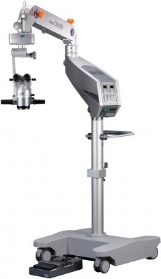
Microscopes | Takagi OM-19 Operating Microscope
The world’s first surgical microscope with independent light intensity adjustment for coaxial and red-reflex illumination to suit each patient case and surgeons unique preference.
High-performance LED surgical microscope combining ‘visualising’ with ‘ease of use’.


Takagi OM-19 Operating Microscope
Tiltable Binocular Tubes
• Tiltable binocular tubes with an incline of more than 90º to suit the height and posture of the operating surgeon.
Movable range: 0º (straight) to 90º (inclined).
• High-grade high-eyepoint multi-coated eyepieces delivering a bright and sharp view.
Easy-to-see Operation Panel
• Light intensity for coaxial and red-reflex illumination can be adjusted separately.
• Focus, zoom, and X-Y speed are independently adjustable.
• Automatic X-Y and focus centering can be performed at the touch of a button.
Foot Controller
• Light intensity for coaxial and red-reflex illumination can be adjusted separately.
• Focus, zoom, and X-Y speed are independently adjustable.
• Automatic X-Y and focus centering can be performed at the touch of a button.
Microscope Head Tilting Mechanism
•By adopting a fine movement interlock system, the microscope head tilts back and forth easily, necessary in the application of glaucoma surgery, with a movable range of around 30º each way.
Safety Stop Mechanism
•Safety stop mechanism equipped as standard, where the lowest arm position can be set according to the height of the operating table, critical during VR procedures.
New Optics
• New red reflex illumination system providing extremely bright reflected light from the fundus, allowing Surgeons to perform cataract surgery with ease.
• By adopting a standard objective lens of F=200 mm, the working distance is optimized for vitreoretinal surgery.
• 10x magnification eyepieces bring a wider, brighter, and sharper view with a larger depth of focus.
Light Source
The Independent LED Light Sources
• Light intensity for red-reflex illumination & coaxial illumination can be adjusted separately due to their fully independent light sources.
High-Illumination LED
• Bright and sharp high-luminance LED having excellent colour balance, giving a much brighter reflected light from the fundus in combination with the new red reflex illumination optical system.es.
Blue Light Reduction
• With the blue correction filter, the projected light is soft and easy on the patient’s eye by reducing the peak of the characteristic blue wavelength of LED.
Assistant Microscope Options
Rotating Coaxial Stereoscopic Assistant’s Microscope
Rotating Coaxial Stereoscopic Assistant’s Microscope
• Stereoscopic view available also through the assistant’s microscope.
• 180˚ continuous rotation through right and left directions whilst mounted.
• Magnification adjusted manually in three steps.
• Tiltable binocular tubes equipped as standard, which can incline more than 90˚.
*Red reflex image through the assistant’s microscope is different than the main surgeons view, the red reflex illumination is optimized for the main surgeon’s optics.
Fixed Coaxial Monoscopic Assistant’s Microscope
• Providing the same coaxial image and red reflex illumination as the main surgeon.
Mounted using the O11-03 Beam splitter, with the optics in/out switching lever, ideal when 100% light is required by the main surgeon.
C-mount Adapter
• CCD camera system can be mounted using O11-03 Beam splitter and O08-22 or O08-11 C-mount camera adaptor for integration with surgical video documentation systems.
• O08-11 is equipped with an adjustable aperture for regulating light, O08-22 comes with a fixed aperture.
Various Options
Monitor Arm & Camera Control Rack
• LCD monitor can be mounted on the upper microscope arm with O06-29 Monitor arm.
• Camera control unit can be mounted on the lower microscope pole with O06-30 Camera control rack.
Fundus Observation Devices Adaptations
• Oculus BIOM and Haag Streit EIBOS adaptations are available.
| Microscope | Magnification Changer | Motorised zoom type (zoom ratio 1:6) | ||
| Objective | F=200mm | |||
| Eyepieces | 10x (high-eyepoint & wide field) | |||
| Binoculars | Tiltable Binocular Tubes F=170mm | |||
| Total magnifications | 3.4x to 20.4x | |||
| Real fields of view | φ58.8mm to φ9.8mm | |||
| Focusing stroke | 50mm (with centering function) | |||
| X-Y movement stroke | ±25mm in each direction (with centering function) | |||
| Illumination | System | Direct illumination | ||
| Light source | LED | |||
| Field of illumination | φ60mm | |||
| Field of red reflex illumination | φ20mm | |||
| Illumination control | 9 steps | |||
| Filters | Heat-absorbing, Blue Correction, Yellow, RetinaShield | |||
| Arm, base | Mount | Floor stand | ||
| Maximum arm extension | 1260mm | |||
| Arm vertical stroke | 500mm | |||
| Base size | 700mm × 740mm | |||
| Other | Weight | 163kg | ||
| Power consumption | 110VA | |||
| Power supply | AC 100-230V, 50/60Hz | |||
Products
Products by type
Diagnostic ImagingOphthalmic Diagnostic Equipment - Aiding Social DistancingOphthalmic LensesSlit Lamps and Ophthalmic LensesTonometry and Visual FieldsSight Testing and RefractionCornealConsultation FurnitureUltrasoundSurgical BedsLasers and ProbesSurgicalConsumablesProducts
Products by brand
Arc Optical LtdBon OpticBovieChongqing Sunkingdom MedicalCorza OphthalmologyESI Inc.EvansInvotecIridexKeelerMeccanotticaMediWorksMurrayNeoLightOculusOptotek MedicalPlusoptixReichertRodenstock InstrumentsSurgistarTakagiUFSKVELAViaLase®VisionixVolkZiemerService
Engineering support Clinic design serviceSpecial Offers
Current offersProduct Focus
IRIDEX Cyclo G6™Reichert® Tono-Vera®ViaLase® ViaLuxe™ LaserMicroPulse®Takagi 700GL LEDUFSK 500XLEVELA ‘Move+’Corza Medical Hands-FreeNeoLight Phoenix ICONPolicies & Certificates
ISO Certificate WEEE Policy GDPR Policy GDPR Auth form Social Responsibility Document Modern Slavery Statement Carbon Reduction Plan Sustainability ReportCarleton Optical
Pattisson House
Addison Road, Chesham
Buckinghamshire HP5 2BD
UK
Email:
carleton@carletonltd.comSales:
01494 775 811
 Subscribe via RSS
 Join us on LinkedIn
 Follow us on Twitter
 Find us on Facebook