


Surgical
Products by type
Diagnostic Imaging
Ophthalmic Diagnostic Equipment - Aiding Social Distancing
Ophthalmic Lenses
Slit Lamps and Ophthalmic Lenses
Tonometry and Visual Fields
Sight Testing and Refraction
Corneal
Consultation Furniture
Ultrasound
Surgical Beds
Lasers and Probes
Consumables
Microscopes | Rodenstock REM 4000 Endothelium Microscope
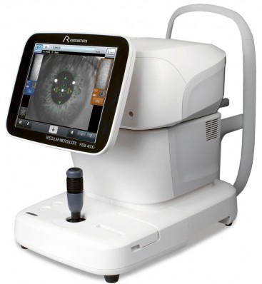
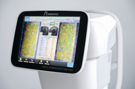
The REM 4000 captures high-quality photos and automatically analyses the size, shape, and population of the endothelial cells. The monitoring of contact lens wearers and evaluation of common ocular conditions become an easy routine.


Rodenstock REM 4000 Endothelium Microscope
- 13 measurement areas
- Counts up to 300 cells
- Integrated non-contact Pachymetry
- Automatic analysis, L-count, Core method, Dark Area method
- Integrated database
- Auto alignment + auto measurement
| Resolution | |
| Pixels used for picture taking: | 480 (V) x 180 (H) pixels |
| Capturing scope: | 0.25 x 0.54 mm |
| 1 center + 12 peripheral measurements: | 13 x fixation points |
| Min. cell resolution: | 1.14 μm (V) x 1.45 μm (H) |
| Optical magnification: | x 190 |
| Display: | 10.4’’ LCD Colour |
| Display resolution: | 1.14 μm |
| Measurement | |
| Auto alignment: | Yes |
| Auto measurement: | Yes |
| Manual mode (1 & 2): | Yes |
| Measurement function | |
| Automated captured examination: | 16 pictures for analysis |
| Number of analysed cells: | Up to 300 cells |
| Capturing position: | Center + 12 peripheral points |
| Analysis method: | Automatic analysis, L-count, Core method, Dark area method |
| Analysis values: | CD (cell density), AVG (average cell area), SD (standard deviation of cell area) |
| CV (coefficient of variation of cell area), Cell size (max. + min. cell area) | |
| Stroke of moving section: | X: 88 mm, Y: 40 mm, Z: 50 mm |
| Stroke of electrical chin rest: | 70 mm |
| Measuring accuracy Pachymetry: | +/- 10 μm |
| Operating Environment | |
| Temperature: | +10° to +40° |
| Humidity: | 30% to 75% |
| Atmospheric pressure: | 800 to 1060 hPa |
| Standards applied: | MDD Annex ii, iSo 13485 |
| Data Management | |
| Built-in printer: | Thermal printer |
| Data output type: | USB-Hx2, USB-Dx2, LAN, SD Card (for internal database) |
| Dimensions & Electric Requirements | |
| Dimensions WDH: | 309 x 491 x 450 mm |
| Weight: | Approx. 22 kg |
| Voltage: | AC 100 to 240 V |
| Frequency: | 50/60 Hz |
| Power consumption: | 100 VA |
Products
Products by type
Diagnostic ImagingOphthalmic Diagnostic Equipment - Aiding Social DistancingOphthalmic LensesSlit Lamps and Ophthalmic LensesTonometry and Visual FieldsSight Testing and RefractionCornealConsultation FurnitureUltrasoundSurgical BedsLasers and ProbesSurgicalConsumablesProducts
Products by brand
Arc Optical LtdBon OpticBovieChongqing Sunkingdom MedicalCorza OphthalmologyESI Inc.EvansInvotecIridexKeelerMeccanotticaMediWorksMurrayNeoLightOculusOptotek MedicalPlusoptixReichertRodenstock InstrumentsSurgistarTakagiUFSKVELAViaLase®VisionixVolkZiemerService
Engineering support Clinic design serviceSpecial Offers
Current offersProduct Focus
IRIDEX Cyclo G6™Reichert® Tono-Vera®ViaLase® ViaLuxe™ LaserMicroPulse®Takagi 700GL LEDUFSK 500XLEVELA ‘Move+’Corza Medical Hands-FreeNeoLight Phoenix ICONPolicies & Certificates
ISO Certificate WEEE Policy GDPR Policy GDPR Auth form Social Responsibility Document Modern Slavery Statement Carbon Reduction Plan Sustainability ReportCarleton Optical
Pattisson House
Addison Road, Chesham
Buckinghamshire HP5 2BD
UK
Email:
carleton@carletonltd.comSales:
01494 775 811
 Subscribe via RSS
 Join us on LinkedIn
 Follow us on Twitter
 Find us on Facebook